Meet the SIRIC Community
💡Meet the people behind SIRIC Montpellier Cancer: Discover inspiring profiles of clinicians, researchers, patient partners, and many others who are committed to advancing our understanding, prevention, and treatment of cancer.
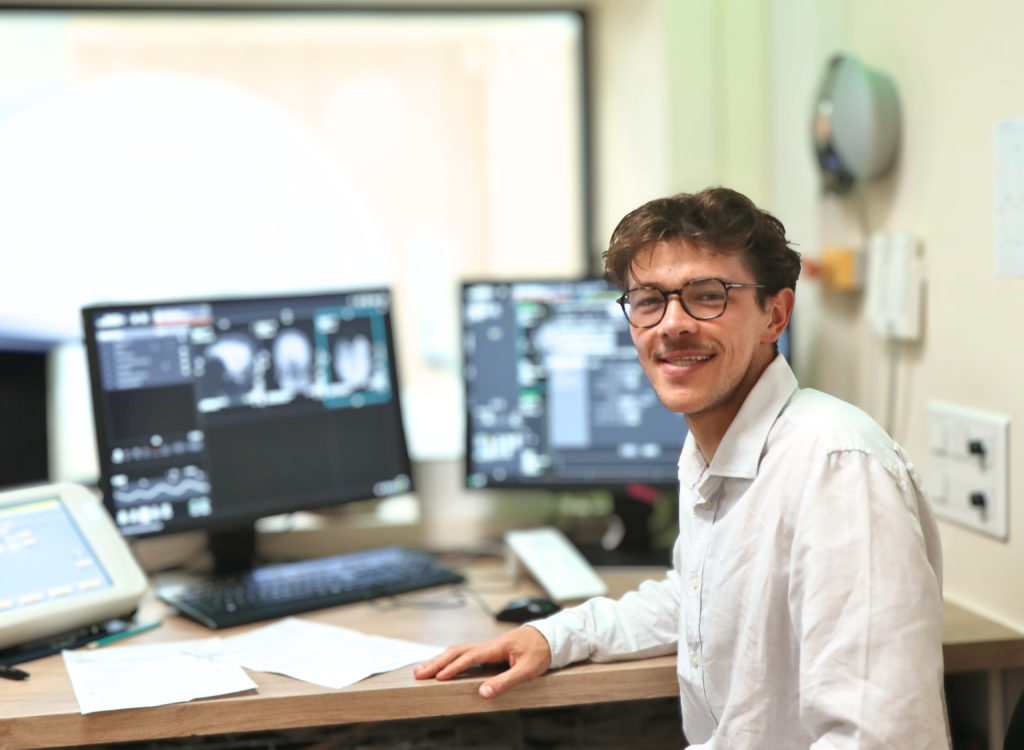
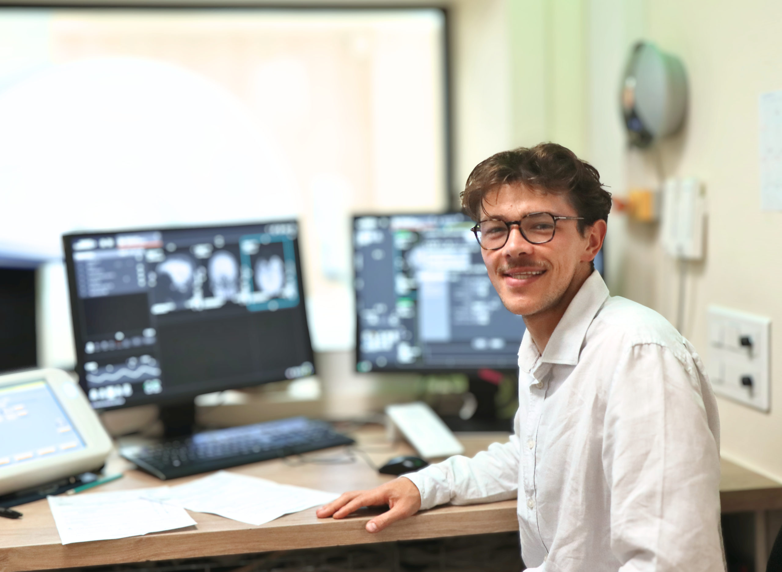
Florent Tachenne, CIFRE PhD Student within the PINKCC Research Team
As part of a CIFRE PhD thesis conducted in collaboration with Siemens Healthineers and the PINKcc research team, led by Professor Stéphanie Nougaret at the IRCM, Florent Tachenne is developing new magnetic resonance imaging (MRI) approaches to better characterize digestive tumors and contribute to more personalized radiotherapy. This work is part of the SIRIC Montpellier Cancer PRIORITY research program.
Trained as a general engineer, Florent chose to apply his skills in physics, mathematics, and programming to medical research through various experiences in both academic laboratories and industry. Today, he tells us about the objectives and challenges of his PhD research.
- What is the clinical issue at the heart of your PhD research?
Today, digestive cancers, particularly pancreatic, liver, and rectal cancers, are still largely managed according to standardized treatment protocols. In radiotherapy, for example, patients generally receive similar radiation doses. However, treatment response can vary considerably from one patient to another. This heterogeneity is partly explained by the specific characteristics of each tumor.
Among the factors that can influence the response to radiotherapy, tumor oxygenation plays an important role. In vitro studies have shown, in particular, that a hypoxic tumor — meaning one that is insufficiently oxygenated — may require up to three times more radiation to achieve a comparable biological effect to that observed in a properly oxygenated tumor.
One of the challenges, therefore, is to identify and characterize these differences as early as possible in the patient care pathway, in order to better tailor treatments to each individual patient.
- What does your work involve, and how do you carry it out?
My work focuses on developing advanced MRI methods to non-invasively identify biomarkers associated with tumor hypoxia. The aim is to obtain this information without making patient care more burdensome, by using an imaging modality that is already integrated into their care pathway.
More specifically, my work involves several stages. I develop and implement new MRI sequences, then test them and perform the acquisitions in patients. I also develop tools to analyze the data collected, particularly using artificial intelligence methods, before interpreting the results obtained.
We are exploring two complementary approaches in particular. The first involves studying tumor acidity. Hypoxic tumors produce more lactate, which can lead to a decrease in tumor pH. Specific quantitative MRI techniques, such as Chemical Exchange Saturation Transfer (CEST), can be used to investigate certain metabolic markers associated with these changes.
The second approach aims to assess tumor oxygenation based on deoxyhemoglobin concentration, using several quantitative MRI sequences.
The objective is then to combine these different sources of information in order to better characterize tumors before radiotherapy begins and identify the parameters that may influence their response to treatment.
- What are the main challenges of your PhD research?
The first challenge is to better understand tumor heterogeneity. Tumors exhibit highly variable characteristics from one patient to another, but also within the same tumor. The goal is therefore to identify relevant tumor profiles and, ultimately, to better understand their relationship with treatment response.
The second challenge is to make MRI measurements sufficiently reliable, sensitive, and specific to be used in different clinical settings. This involves developing robust and reproducible analytical methods, while also taking into account variations that may exist between different imaging systems and acquisition protocols. Artificial intelligence may, in particular, help improve the robustness of these analyses.
Finally, the longer-term goal is to develop tools that can be used in clinical practice and on a large scale. This requires working with sufficiently large cohorts to validate the approaches developed and determine their clinical relevance.
This PhD research therefore aims to bridge methodological developments in medical imaging, data analysis, and their potential translation into clinical practice, with the ambition of contributing to radiotherapy that is better adapted to the specific characteristics of each tumor.